

Hypoxic Conditioning: When Less (Oxygen) is More
Alex Roth
Illustrations by Anna Bishop

What comes to mind when you think of the mountains? Is it hiking? Snow? Bighorn sheep? Alzheimer’s Disease…? Alzheimer's Disease (AD) might not be the first thing that comes to mind when you think of the mountains. However, AD-related deaths have been shown to decrease as altitude increases and, correspondingly, air pressure and oxygen levels decrease [1]. At first glance, there is no obvious link between altitude and AD mortality, but what if the connection between lowered air pressure and AD could be revolutionary in the management of this disease that affects 30 million people worldwide [2]? Enter hypoxic conditioning: an emerging preventative treatment for a number of conditions including neurodegenerative disease, stroke, and heart attack [3, 4, 5]. Hypoxic conditioning (HC) is a treatment that trains you to resist lowered oxygen levels in your body, a state called hypoxia. Experiencing hypoxia causes the body to react by trying to obtain more oxygen and defaulting to other ways to make energy, which may make one feel lightheaded or groggy. Like a vaccine that introduces small amounts of a disease to train your immune system's resistance, HC deprives you of oxygen for short periods of time to train your body to withstand hypoxia. Over time, this reaction results in a vaccine-like resistance to the sustained hypoxia seen in many diseases related to aging.
AD is part of a group of neurological illnesses known as neurodegenerative diseases, which cause deterioration and death in the cells of the brain and spinal cord. This degeneration causes dementia, which is classified as the loss of multiple mental functions including memory, reasoning, normal personality, and language [6]. AD is the most common cause of dementia, which can appear subtly over many years and be extremely debilitating [2]. AD is the sixth leading cause of death worldwide, largely due to ineffective treatment options that fail to address the full scope of the disease [2, 7].
Your Brain on Hypoxia
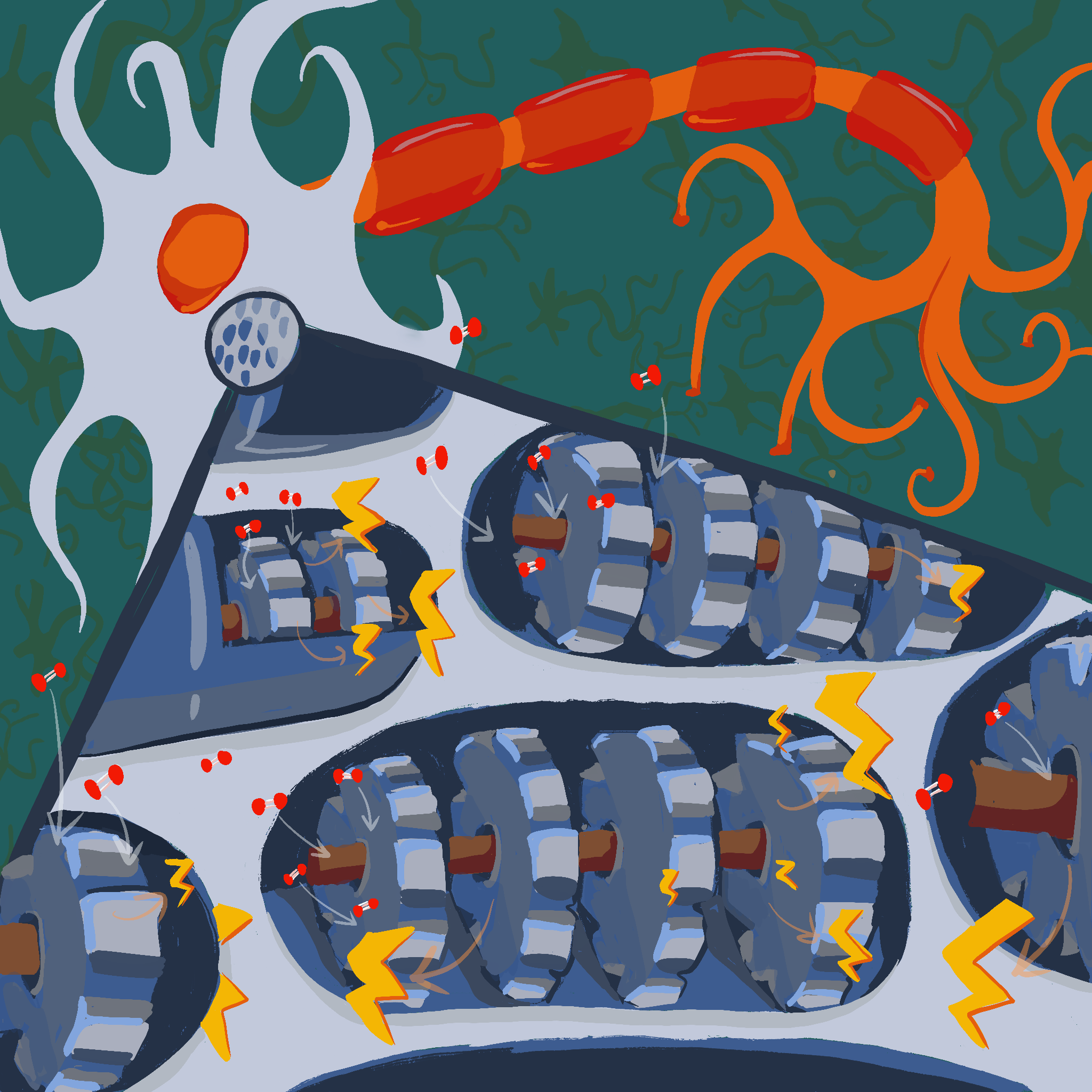
To understand how diseases like AD affect us, we must first understand how our brain’s metabolism works. The keystone of our body’s energy production is the mitochondrion, often known as the ‘powerhouse of the cell.’ The mitochondrion converts the body’s fuel — glucose — to energy, and stores it as adenosine triphosphate (ATP), the universal energy currency for biological systems. The mitochondria can be thought of as cellular engines, since they use a constant flow of fuel and oxygen to maintain ATP production rates. Your body usually generates energy using oxygen through a pathway called aerobic respiration [8]. However, even if you are breathing enough oxygen from the air around you, it may not be properly distributed to different areas of your body, causing hypoxia in these areas. Hypoxia forces the body into a different type of respiration: anaerobic respiration. Though it uses no oxygen, this method of energy production generates substantially fewer ATP per glucose molecule [5]. Furthermore, anaerobic respiration results in the overproduction of mitochondrial byproducts called reactive oxygen species (ROS), which damage cell structures and processes. ROS molecules can cause neuroinflammation, the brain’s ‘scorched earth’ immune response against the damaged cellular architecture that results in widespread damage to the brain. If your brain is forced into anaerobic metabolism for too long, neurodegeneration and cell death begin to occur [5].
The brain requires large amounts of energy to function effectively: it uses one fifth of all the oxygen in your body and one fourth of the glucose. When there is plenty of oxygen present, it prefers the greater efficiency of aerobic metabolism [8, 9]. In some instances, however, our bodies lack the oxygen required to undergo aerobic metabolism. Following a period of intense exercise, your brain enters a state of hypoxia. As a result, you may feel lightheaded, confused, tired, anxious, uncoordinated, and clumsy [10, 11]. These sudden changes are a result of your brain failing to keep up with its energy production needs, causing cognitive function to temporarily deteriorate. Hypoxia is particularly dangerous for the cells of the brain, or neurons, because they have higher energy requirements than other cells [5]. Fundamentally, the body needs two main ingredients for sustained energy production: glucose and oxygen. However, during hypoxia, there is not enough oxygen for sustained aerobic metabolism, so the body switches to anaerobic metabolism.

A Workout for Your Brain: the Principles of Hypoxic Conditioning
Since the body prefers anaerobic metabolism in hypoxic conditions, the idea of HC might seem perplexing [8]. How could repeated exposure to a dangerous state be safe, let alone helpful? Let’s take a look. Like athletic conditioning, HC uses short ‘workouts’ for your metabolism, which activate hypoxic defenses against diseases like AD. Over time, these ‘workouts’ help the body endure low oxygen levels. Just as a runner jogs every day to prepare for a marathon, HC would expose people to controlled periods of hypoxia, conditioning them against metabolic disease [12].
A marathon runner on their morning jog needs more blood pumping to their leg muscles than if they were resting, so their heart rate may increase for the duration of their run. Similarly, our brains have evolved a number of short-term defenses against oxygen deprivation, which have the potential to be advantageous when implementing HC [13]. Without these defenses, brain cells would quickly lose function and eventually die. One such defense is an initial upregulation of aerobic metabolism, which sustains the brain by generating as much ATP as possible while some oxygen still remains [14, 15, 16]. The brain also increases anaerobic metabolism as a backup strategy to continue producing a small amount of energy [15]. A number of proteins that modulate our body’s responses to hypoxia, termed hypoxia inducible factors (HIFs), are activated as well [5]. Activation of HIFs ‘turns on’ antioxidant molecules that help neutralize ROS byproducts by dampening their ability to damage and inflame the brain. Finally, the brain increases its glucose intake to refuel its cells [17]. The promise of HC lies in how it leverages these defenses. Instead of simply activating these mechanisms for short-term protection, HC promotes the development of a long-term protective state that is resistant to hypoxic damages [13].
Once again, imagine our marathon runner: they may not see immediate improvements in their running, but over time their leg muscles, heart, and lungs will strengthen and grow to support their body as they run. Similarly, repeated bouts of HC help the brain become more resilient in the face of long-term oxygen deprivation, decreasing potential damage to brain cells. HC stimulates new growth of blood vessels, enabling future increases in blood flow and thereby supporting glucose and oxygen delivery to the brain [17]. The brief disturbances HC causes in the brain’s oxygen levels also train the brain to better resist disruption of ATP generation [18, 19]. Finally, HC activates protective antioxidant mechanisms — including HIFs — against long-term ROS production [20]. Activating these defenses helps prevent the damages that hypoxia and subsequent decreased metabolism can cause [21, 22, 23]. Now, under the duress of hypoxia, the brain can rely on not only its standard defenses, but also its newfound resilience resulting from HC. However, HC does have the potential to damage the brain [24]. Just as runners must be careful to avoid injuries from overtraining, the duration of hypoxia needs to be carefully controlled during HC to keep the brain safe from injury [22, 25]. Fortunately, oxygen can be delivered at intervals that both induce the benefits of HC and keep the patient safe [22, 25]. In doing so, healthcare providers can take advantage of HC to help the rapidly growing population at risk of AD [26].
Into Thin Air: Treating Alzheimer’s Disease with Hypoxic Conditioning
Much like our bodies, our brains also slow and decay with age. In the process, metabolism typically becomes less efficient, which — along with other processes — results in neurodegeneration [27]. The same metabolic alterations responsible for the aging of the brain are also seen in a more advanced form of AD [28, 29]. Decreased blood flow to the brain is thought to be the earliest manifestation of AD [30]. Soon after, mitochondrial and metabolic processes begin to malfunction, hindering the brain’s ability to produce energy [31, 32]. In turn, these malfunctions lead to chronic hypoxia and lowered brain metabolism, both of which are major contributors to the symptoms of AD [33, 34, 35]. Fortunately, the possibility of utilizing HC to decrease the metabolic dysfunction of AD is currently being explored [21, 36].
Though AD has significant non-metabolic components, HC mainly shows promise in its ability to mitigate metabolic deficits [12]. Reduced blood flow to the brain can be used to predict behavioral symptoms like memory difficulties before they emerge [9, 37]. Brain cells in people with AD typically have lower metabolic rates than healthy brain cells, potentially because less oxygen and glucose are being delivered to them through the blood [9, 38]. As a result, brains are forced to rely more heavily on anaerobic metabolism, resulting in damage to brain cells from excessive ROS generation [22, 38]. Remember HIFs — the proteins HC activates? HC can keep HIFs activated for a long time and provide a safe way to activate them before they are needed [5, 39]. When activated by HC, HIFs express red blood cell and blood vessel production genes, thereby increasing oxygen-rich blood delivery to the brain [17, 35]. The addition of new blood cells and vessels is akin to adding more delivery trucks to a mail route; now more oxygen-rich blood can be delivered to brain cells for aerobic metabolism.
Like running a dryer filled with lint, mitochondria start to wear under the neuroinflammatory stress of AD [36]. Malfunctioning mitochondria are a significant metabolic aspect of AD [31, 40, 41]. Mitochondria are essential in generating the ATP needed for brain survival and function. Therefore, processes such as speech, memorization, and planning are all impaired by lowered mitochondrial function, because fewer ATP molecules are available to power the communication between neurons [42, 43]. HC fortunately shows promise in alleviating mitochondrial dysfunction, as it can stabilize malfunctioning mitochondria and increase growth in lab-grown cells [18, 35]. Furthermore, mitochondria in hypoxically conditioned rats exhibit elevated antioxidant activity and resistance to hypoxia [35]. Perhaps most surprisingly, HC was able to drastically improve survival of mice with experimentally induced mitochondrial disease [18]. Though all untreated mice died by the second month, mice treated with HC only showed mild symptoms by their fourth month [18]! Even though animal results cannot be directly translated to humans, HC appears to limit the effects of mitochondrial disease and dysfunction in model organisms, suggesting a promising future for use in human treatment.
Overproduction of ROS is another major stressor implicated in AD [44]. In a rodent model of AD, ROS were shown to play a role in neuroinflammation and cell death [22]. As a result, neurons exposed to ROS were damaged by the brain’s immune system, leading to cognitive deficits such as impaired memory [22]. Just as a vaccine introduces a weakened version of a virus, the brief hypoxic episodes utilized in HC caused low-level generation of ROS [21]. ROS induce a response that, with reinforcement from multiple phases of conditioning, introduces long-term resistance to ROS damage [21]. The hypoxic ‘vaccine’ effect likely helps to reduce neuroinflammation by releasing antioxidant molecules and activating other neuroprotective systems [20, 23]. Essentially, HC trains the brain to more efficiently neutralize ROS produced by AD before they can cause neurodegeneration. However, AD is a remarkably complex disease with metabolic and non-metabolic components. Thus, the extent to which HC can act as an effective therapeutic at all stages of AD will remain unknown until human trials occur.
One way AD damage can manifest is in the loss of connections between neurons, which prevents signals from traveling through the brain [32]. Brain cells become vulnerable to damage and death when starved of ATP because they cannot keep up with energetic demands for maintenance and function [27]. In fact, metabolic dysfunction is widely considered to be one of the key factors involved in AD neurodegeneration, as neurons in areas of the brain associated with memory waste away [9, 45, 46]. The goal of HC is to provide a cheap and effective way to restore metabolic and cognitive function, and apply rodent models to human trials to explore its potential as a treatment for sick people in the future. However, questions remain to be addressed before HC becomes a reality.

One of the most prominent questions which must be confronted before HC can be implemented is what exactly constitutes a safe level of treatment [12, 47]. While experiments with animals suggest that there are safe levels of hypoxia to use in conditioning, human trials are still needed to assess HC’s safety and efficacy [22, 25]. Continued research may enable the testing and assessment of both preventative and therapeutic HC treatment in humans for a wide variety of diseases, not just AD [12]. Luckily, HC shows great potential for future advances since it is a novel therapy for neurodegeneration that does not involve expensive, inaccessible medications [48]. As a technique, rather than a physical product, HC could be practiced globally without the need for continuous production and shipping of expirable medication. All these factors may help soften the blow Alzheimer’s disease inflicts on a growing proportion of the elderly population [2].
References
1. Thielke, S., Slatore, C. G., & Banks, W. A. (2015). Association between Alzheimer dementia mortality rate and altitude in California counties. JAMA Psychiatry, 72(12), 1253–1254. doi:10.1001/jamapsychiatry.2015.1852.
2. Haque, R. U., & Levey, A. I. (2019). Alzheimer’s disease: a clinical perspective and future nonhuman primate research opportunities. Proceedings of the National Academy of Sciences, 116(52), 26224–26229. doi:10.1073/pnas.1912954116.
3. Mallet, R. T., Manukhina, E. B., Ruelas, S. S., Caffrey, J. L., & Downey, H. F. (2018). Cardioprotection by intermittent hypoxia conditioning: evidence, mechanisms, and therapeutic potential. American Journal of Physiology-Heart and Circulatory Physiology, 315(2), H216–H232. doi:10.1152/ajpheart.00060.2018.
4. Rhudy, J. P., Bakitas, M. A., Hyrkäs, K., Jablonski‐Jaudon, R. A., Pryor, E. R., Wang, H. E., & Alexandrov, A. W. (2015). Effectiveness of regionalized systems for stroke and myocardial infarction. Brain and Behavior, 5(10), e00398. doi:10.1002/brb3.398.
5. Mitroshina, E. V., Savyuk, M. O., Ponimaskin, E., & Vedunova, M. V. (2021). Hypoxia-inducible factor (HIF) in ischemic stroke and neurodegenerative disease. Frontiers in Cell and Developmental Biology, 9, 703084. doi:10.3389/fcell.2021.703084.
6. Weller, J., & Budson, A. (2018). Current understanding of Alzheimer’s disease diagnosis and treatment. F1000Research, 7, 1161. doi:10.12688/f1000research.14506.1.
7. Yiannopoulou, K. G., & Papageorgiou, S. G. (2020). Current and future treatments in Alzheimer disease: an update. Journal of Central Nervous System Disease, 12. doi:10.1177/1179573520907397.
8. Magistretti, P. J., & Allaman, I. (2013). Brain energy metabolism. In D. W. Pfaff (Ed.), Neuroscience in the 21st Century: From Basic to Clinical, 1591-1620. doi:10.1007/978-1-4614-1997-6_56.
9. Watts, M. E., Pocock, R., & Claudianos, C. (2018). Brain energy and oxygen metabolism: emerging role in normal function and disease. Frontiers in Molecular Neuroscience, 11, 216. doi:10.3389/fnmol.2018.00216.
10. Neuhaus, C., & Hinkelbein, J. (2014). Cognitive responses to hypobaric hypoxia: implications for aviation training. Psychology Research and Behavior Management, 7, 297–302. doi:10.2147/PRBM.S51844.
11. Woodrow, A. D., Webb, J. T., & Wier, G. S. (2011). Recollection of hypoxia symptoms between training events. Aviation, Space, and Environmental Medicine, 82(12), 1143–1147. doi:10.3357/asem.2987.2011.
12. Verges, S., Chacaroun, S., Godin-Ribuot, D., & Baillieul, S. (2015). Hypoxic conditioning as a new therapeutic modality. Frontiers in Pediatrics, 3. doi:10.3389/fped.2015.00058.
13. Rybnikova, E., & Samoilov, M. (2015). Current insights into the molecular mechanisms of hypoxic pre- and postconditioning using hypobaric hypoxia. Frontiers in Neuroscience, 9, 388. doi:10.3389/fnins.2015.00388.
14. Smith, Z. M., Krizay, E., Guo, J., Shin, D. D., Scadeng, M., & Dubowitz, D. J. (2013). Sustained high-altitude hypoxia increases cerebral oxygen metabolism. Journal of Applied Physiology, 114(1), 11–18. doi:10.1152/japplphysiol.00703.2012.
15. Vestergaard, M. B., Lindberg, U., Aachmann-Andersen, N. J., Lisbjerg, K., Christensen, S. J., Law, I., Rasmussen, P., Olsen, N. V., & Larsson, H. B. (2016). Acute hypoxia increases the cerebral metabolic rate – a magnetic resonance imaging study. Journal of Cerebral Blood Flow & Metabolism, 36(6), 1046–1058. doi:10.1177/0271678X15606460.
16. Xu, F., Liu, P., Pascual, J. M., Xiao, G., & Lu, H. (2012). Effect of hypoxia and hyperoxia on cerebral blood flow, blood oxygenation, and oxidative metabolism. Journal of Cerebral Blood Flow & Metabolism, 32(10), 1909–1918. doi:10.1038/jcbfm.2012.93.
17. Merelli, A., Rodríguez, J. C. G., Folch, J., Regueiro, M. R., Camins, A., & Lazarowski, A. (2018). Understanding the role of hypoxia inducible factor during neuro-degeneration for new therapeutics opportunities. Current Neuropharmacology, 16(10), 1484–1498. doi:10.2174/1570159X16666180110130253.
18. Jain, I. H., Zazzeron, L., Goli, R., Alexa, K., Schatzman-Bone, S., Dhillon, H., Goldberger, O., Peng, J., Shalem, O., Sanjana, N. E., Zhang, F., Goessling, W., Zapol, W. M., & Mootha, V. K. (2016). Hypoxia as a therapy for mitochondrial disease. Science, 352(6281), 54–61. doi:10.1126/science.aad9642.
19. Liu, M., & Alkayed, N. J. (2005). Hypoxic preconditioning and tolerance via hypoxia inducible factor (HIF) 1α-linked induction of P450 2C11 epoxygenase in astrocytes. Journal of Cerebral Blood Flow & Metabolism, 25(8), 939–948. doi:10.1038/sj.jcbfm.9600085.
20. Millán, I., Corral-Debrisky, M., Vento, M., & Torres-Cuevas, I. (2022). Hypoxic preconditioning induces neuroprotection against oxidative stress. Redox Experimental Medicine, 2022(1), R159–R167. doi:10.1530/REM-22-0011.
21. Jung, M. E., Simpkins, J. W., Wilson, A. M., Downey, H. F., & Mallet, R. T. (2008). Intermittent hypoxia conditioning prevents behavioral deficit and brain oxidative stress in ethanol-withdrawn rats. Journal of Applied Physiology, 105(2), 510–517. doi:10.1152/japplphysiol.90317.2008.
22. Manukhina, E. B., Goryacheva, A. V., Barskov, I. V., Viktorov, I. V., Guseva, A. A., Pshennikova, M. G., Khomenko, I. P., Mashina, S. Yu., Pokidyshev, D. A., & Malyshev, I. Yu. (2010). Prevention of neurodegenerative damage to the brain in rats in experimental Alzheimer’s disease by adaptation to hypoxia. Neuroscience and Behavioral Physiology, 40(7), 737–743. doi:10.1007/s11055-010-9320-6.
23. Shu, L., Wang, C., Wang, J., Zhang, Y., Zhang, X., Yang, Y., Zhuo, J., & Liu, J. (2016). The neuroprotection of hypoxic preconditioning on rat brain against traumatic brain injury by up-regulated transcription factor Nrf2 and HO-1 expression. Neuroscience Letters, 611, 74–80. doi:10.1016/j.neulet.2015.11.012.
24. Biswal, S., Sharma, D., Kumar, K., Nag, T. C., Barhwal, K., Hota, S. K., & Kumar, B. (2016). Global hypoxia induced impairment in learning and spatial memory is associated with precocious hippocampal aging. Neurobiology of Learning and Memory, 133, 157–170. doi:10.1016/j.nlm.2016.05.011.
25. Stowe, A. M., Altay, T., Freie, A. B., & Gidday, J. M. (2011). Repetitive hypoxia extends endogenous neurovascular protection for stroke. Annals of Neurology, 69(6), 975–985. doi:10.1002/ana.22367.
26. Brookmeyer, R., Johnson, E., Ziegler-Graham, K., & Arrighi, H. M. (2007). Forecasting the global burden of Alzheimer’s disease. Alzheimer’s & Dementia, 3(3), 186–191. doi:10.1016/j.jAlz.2007.04.381.
27. Camandola, S., & Mattson, M. P. (2017). Brain metabolism in health, aging, and neurodegeneration. The EMBO Journal, 36(11), 1474–1492. doi:10.15252/embj.201695810.
28. Denver, P., & McClean, P. L. (2018). Distinguishing normal brain aging from the development of Alzheimer’s disease: inflammation, insulin signaling and cognition. Neural Regeneration Research, 13(10), 1719–1730. doi:10.4103/1673-5374.238608.
29. Leparulo, A., Bisio, M., Redolfi, N., Pozzan, T., Vassanelli, S., & Fasolato, C. (2022). Accelerated aging characterizes the early stage of Alzheimer’s disease. Cells, 11(2), 238. doi:10.3390/cells11020238.
30. Korte, N., Nortley, R., & Attwell, D. (2020). Cerebral blood flow decrease as an early pathological mechanism in Alzheimer’s disease. Acta Neuropathologica, 140(6), 793–810. doi:10.1007/s00401-020-02215-w.
31. Cardoso, S., Carvalho, C., Correia, S. C., Seiça, R. M., & Moreira, P. I. (2016). Alzheimer’s disease: from mitochondrial perturbations to mitochondrial medicine. Brain Pathology, 26(5), 632–647. doi:10.1111/bpa.12402.
32. Tang, J., Oliveros, A., & Jang, M.-H. (2019). Dysfunctional mitochondrial bioenergetics and synaptic degeneration in Alzheimer disease. International Neurourology Journal, 23(Suppl 1), S5-S10. doi:10.5213/inj.1938036.018.
33. Rybnikova, E., Vataeva, L., Tyulkova, E., Gluschenko, T., Otellin, V., Pelto-Huikko, M., & Samoilov, M. O. (2005). Mild hypoxia preconditioning prevents impairment of passive avoidance learning and suppression of brain NGFI-A expression induced by severe hypoxia. Behavioural Brain Research, 160(1), 107–114. doi:10.1016/j.bbr.2004.11.023.
34. Rybnikova, E. A., Samoilov, M. O., Mironova, V. I., Tyul’kova, E. I., Pivina, S. G., Vataeva, L. A., Ordyan, N. É., Abritalin, E. Yu., & Kolchev, A. I. (2008). The possible use of hypoxic preconditioning for the prophylaxis of post-stress depressive episodes. Neuroscience and Behavioral Physiology, 38(7), 721–726. doi:10.1007/s11055-008-9038-x.
35. Manukhina, E. B., Downey, H. F., Shi, X., & Mallet, R. T. (2016). Intermittent hypoxia training protects cerebrovascular function in Alzheimer’s disease. Experimental Biology and Medicine, 241(12), 1351–1363. doi:10.1177/1535370216649060.
36. Correia, S. C., Machado, N. J., Alves, M. G., Oliveira, P. F., & Moreira, P. I. (2021). Intermittent hypoxic conditioning rescues cognition and mitochondrial bioenergetic profile in the triple transgenic mouse model of Alzheimer’s disease. International Journal of Molecular Sciences, 22(1), 461. doi:10.3390/ijms22010461.
37. Roher, A. E., Debbins, J. P., Malek-Ahmadi, M., Chen, K., Pipe, J. G., Maze, S., Belden, C., Maarouf, C. L., Thiyyagura, P., Mo, H., Hunter, J. M., Kokjohn, T. A., Walker, D. G., Kruchowsky, J. C., Belohlavek, M., Sabbagh, M. N., & Beach, T. G. (2012). Cerebral blood flow in Alzheimer’s disease. Vascular Health and Risk Management, 8, 599–611. doi:10.2147/VHRM.S34874.
38. Liguori, C., Chiaravalloti, A., Sancesario, G., Stefani, A., Sancesario, G. M., Mercuri, N. B., Schillaci, O., & Pierantozzi, M. (2016). Cerebrospinal fluid lactate levels and brain [18F]FDG PET hypometabolism within the default mode network in Alzheimer’s disease. European Journal of Nuclear Medicine and Molecular Imaging, 43(11), 2040–2049. doi:10.1007/s00259-016-3417-2.
39. Yuan, G., Nanduri, J., Khan, S., Semenza, G. L., & Prabhakar, N. R. (2008). Induction of HIF-1α expression by intermittent hypoxia: involvement of NADPH oxidase, Ca2+ signaling, prolyl hydroxylases, and mTOR. Journal of Cellular Physiology, 217(3), 674–685. doi:10.1002/jcp.21537.
40. Cai, Q., & Tammineni, P. (2017). Mitochondrial aspects of synaptic dysfunction in Alzheimer’s disease. Journal of Alzheimer’s Disease, 57(4), 1087–1103. doi:10.3233/JAD-160726.
41. Koopman, W. J. H., Willems, P. H. G. M., & Smeitink, J. A. M. (2012). Monogenic mitochondrial disorders. New England Journal of Medicine, 366(12), 1132–1141. doi:10.1056/NEJMra1012478.
42. Norat, P., Soldozy, S., Sokolowski, J. D., Gorick, C. M., Kumar, J. S., Chae, Y., Yağmurlu, K., Prada, F., Walker, M., Levitt, M. R., Price, R. J., Tvrdik, P., & Kalani, M. Y. S. (2020). Mitochondrial dysfunction in neurological disorders: exploring mitochondrial transplantation. NPJ Regenerative Medicine, 5(1), Article 1. doi:10.1038/s41536-020-00107-x.
43. Zhao, X.-Y., Lu, M.-H., Yuan, D.-J., Xu, D.-E., Yao, P.-P., Ji, W.-L., Chen, H., Liu, W.-L., Yan, C.-X., Xia, Y.-Y., Li, S., Tao, J., & Ma, Q.-H. (2019). Mitochondrial dysfunction in neural injury. Frontiers in Neuroscience, 13, 30. doi:10.3389/fnins.2019.00030.
44. Huang, W.-J., Zhang, X., & Chen, W.-W. (2016). Role of oxidative stress in Alzheimer’s disease. Biomedical Reports, 4(5), 519–522. doi:10.3892/br.2016.630.
45. Johnson, K. A., Fox, N. C., Sperling, R. A., & Klunk, W. E. (2012). Brain imaging in Alzheimer disease. Cold Spring Harbor Perspectives in Medicine, 2(4). doi:10.1101/cshperspect.a006213.
46. Zhang, F., Niu, L., Li, S., & Le, W. (2019). Pathological impacts of chronic hypoxia on Alzheimer’s disease. ACS Chemical Neuroscience, 10(2), 902–909. doi:10.1021/acschemneuro.8b00442.
47. Sprick, J. D., Mallet, R. T., Przyklenk, K., & Rickards, C. A. (2019). Ischaemic and hypoxic conditioning: potential for protection of vital organs. Experimental Physiology, 104(3), 278–294. doi:10.1113/EP087122.
48. Rybnikova, E. A., Nalivaeva, N. N., Zenko, M. Y., & Baranova, K. A. (2022). Intermittent hypoxic training as an effective tool for increasing the adaptive potential, endurance and working capacity of the brain. Frontiers in Neuroscience, 16. doi:10.3389/fnins.2022.941740.